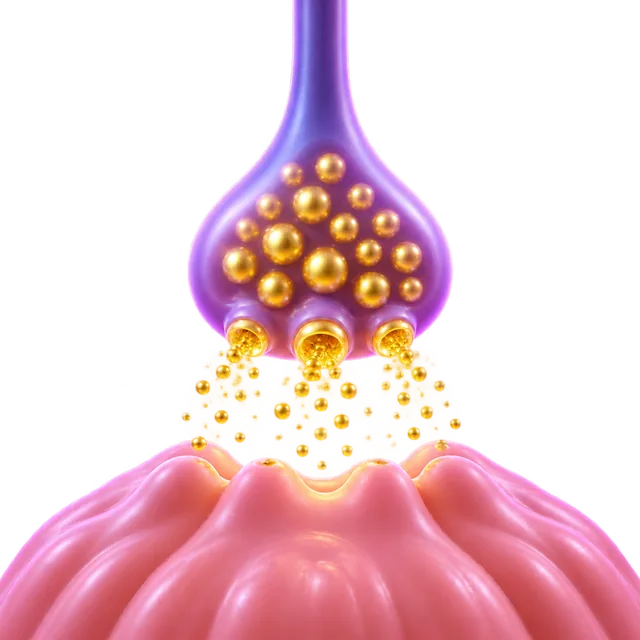
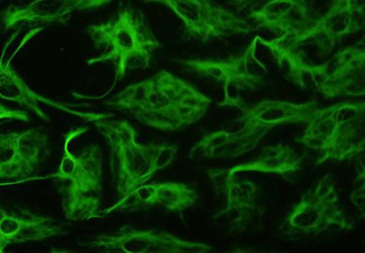
A synaptic vesicle is a microscopic lipid bubble inside a nerve cell that stores chemical messengers called neurotransmitters. When an electrical signal reaches the nerve ending, these tiny spheres fuse with the outer cell membrane to release their contents to neighboring cells. In the human visual cortex, they average just 39.5 nanometers across.
By the edgi team We find the most surprising true thing about an idea and build a 60-second lesson around it.
A neuron's electrical signal cannot jump to the next neuron. There is a tiny gap, the synapse, so the message has to be converted into chemistry. The chemistry is stored in synaptic vesicles: microscopic fat-walled bubbles packed with neurotransmitters, the cell's chemical messengers.
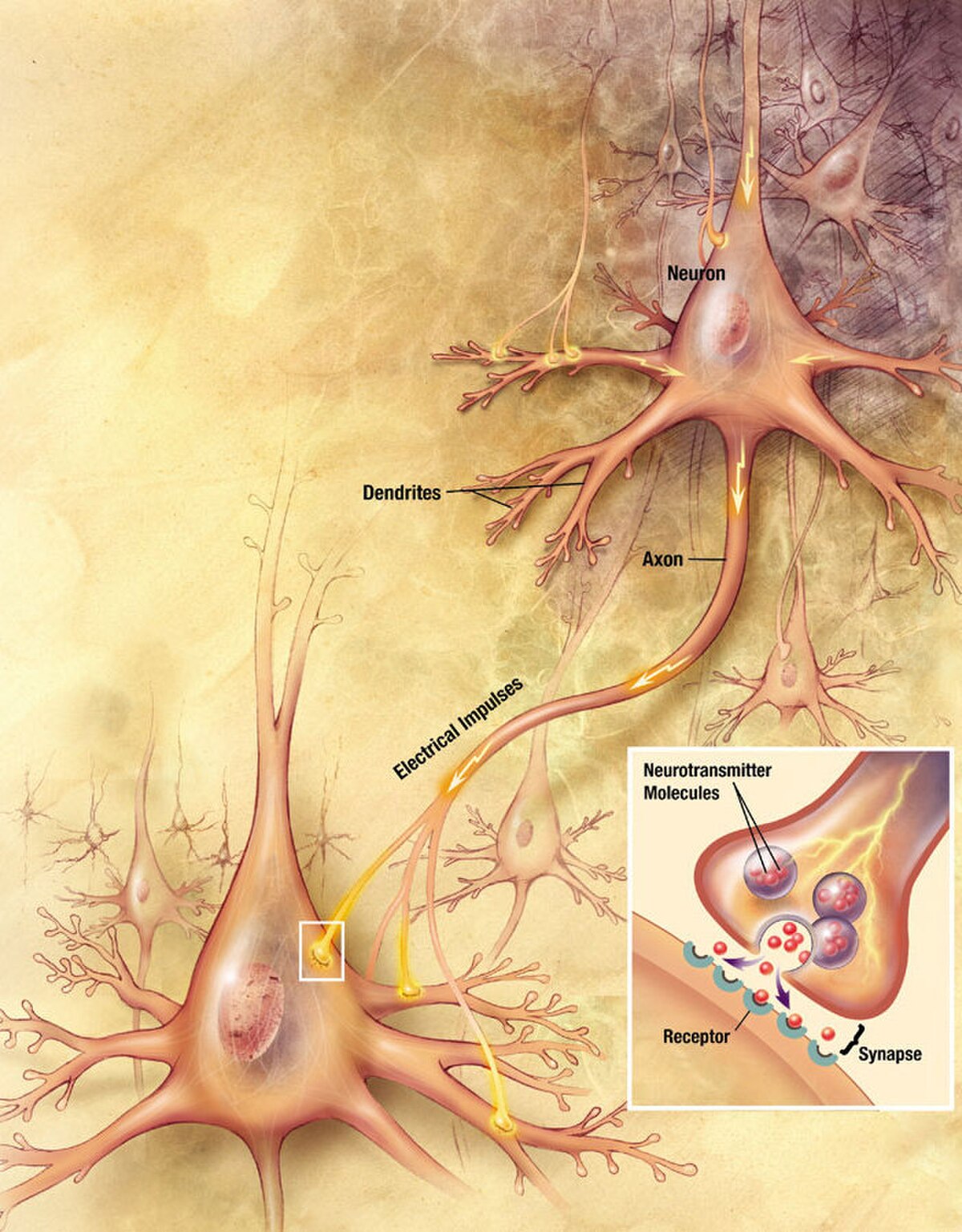
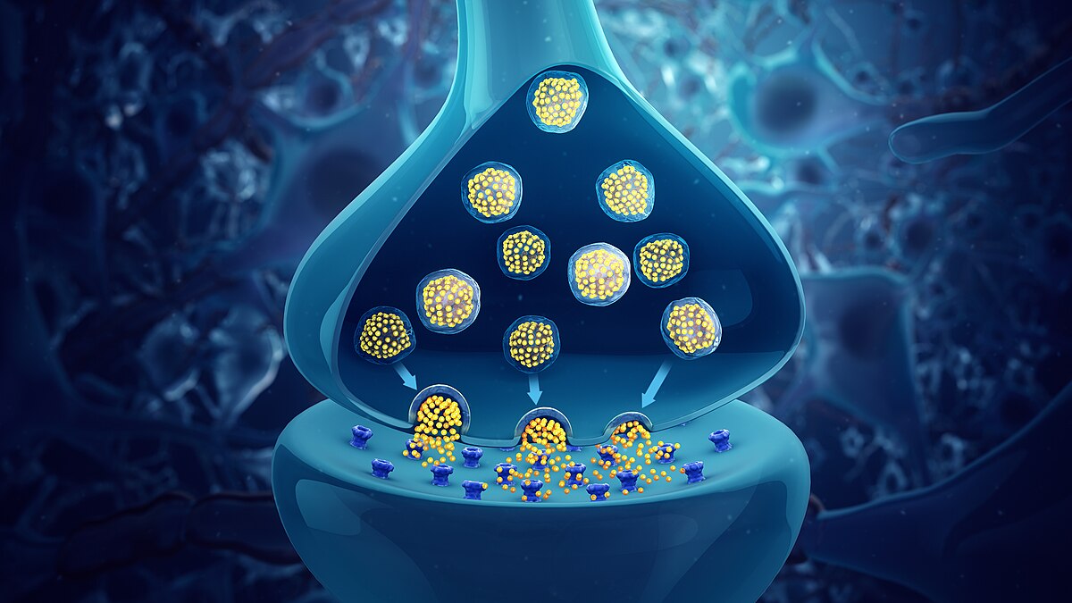
Illustration of a chemical synapse, detailing the process of synaptic transmission between neurons. Illustrator: Christy Krames, MA, CMI, for US National Institutes of Health, National Institute on Aging, Public domain, via Wikimedia Commons
A single vesicle is only about 40 nanometers wide, and a nerve ending holds hundreds of them clustered near its edge, like loaded cartridges.
The trigger
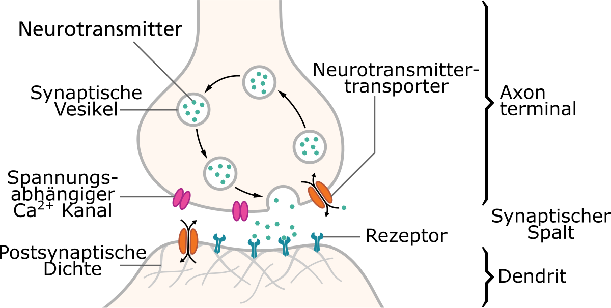
When an electrical pulse reaches the nerve ending, it opens calcium channels, gates that respond to voltage. Calcium floods in. Calcium is the trigger. It makes docked vesicles fuse with the cell membrane and dump their neurotransmitters into the gap, in a fraction of a millisecond.
Diagram of a synapse in German, illustrating the release and reuptake of neurotransmitters. Thomas Splettstoesser (www.scistyle.com), CC BY-SA 4.0, via Wikimedia Commons
The messengers drift across, bind to the next cell, and pass the signal on. Then the cell clears them away, partly by reuptake, pulling them back in, to end the message.
The recycling
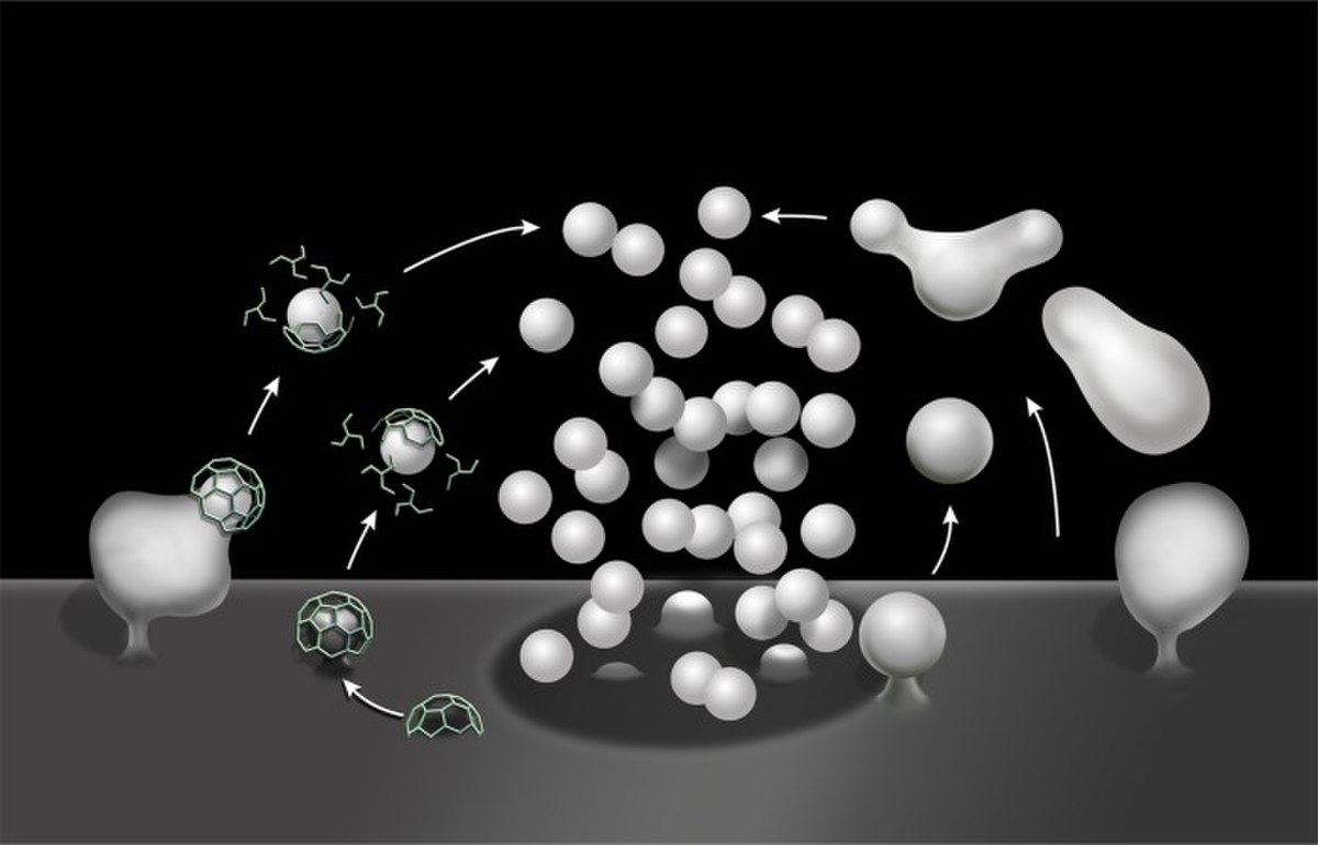
A neuron can't afford to run out. After releasing, the cell pulls the vesicle membrane back inside, rebuilds the bubble, and refills it using transporter proteins that pump in neurotransmitter. Even so, a terminal releases only a limited number: up to about 130 vesicles per ending over ten minutes of slow stimulation.
Schematic drawing illustrating clathrin-mediated (left) and clathrin-independent endocytosis (right) of synaptic vesicle membranes. Wu Y, O'Toole E, Girard M, Ritter B, Messa M, Liu X, McPherson P, Ferguson S, De Camilli P, CC BY 4.0, via Wikimedia Commons
The machinery is fragile in a useful way. Botulinum toxin, the poison behind Botox, snips the proteins vesicles need to fuse. No release means muscles get no signal and go slack.
How synaptic vesicles load and release signals
A synaptic vesicle relies on a specialized proton pump called V-ATPase. This pump burns ATP for energy to create an electrochemical gradient, driving transport proteins to pump neurotransmitters like glutamate, acetylcholine, or GABA out of the cytoplasm and into the vesicle.
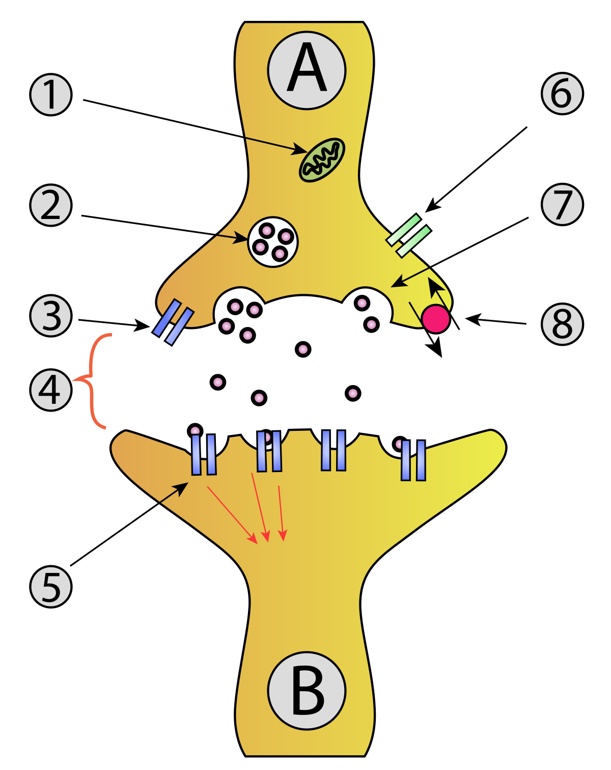
Synaptic vesicles (labeled 2) cluster at the presynaptic axon terminal ready to release chemical signals across to the receiving neuron. vectorization: Mouagip (talk) Synapse_diag1.png: Drawn by fr:Utilisateur:Dake Corrections of original PNG by en:User:Nre, CC BY-SA 3.0, via Wikimedia Commons
After loading, the vesicle travels through distinct steps: docking near the cell membrane, priming, and fusion. During priming, proteins like Munc13 and RIM help assemble SNARE protein complexes. When calcium floods into the cell through voltage-dependent channels, the calcium-sensing protein synaptotagmin triggers the SNAREs to complete membrane fusion, dumping the neurotransmitters out.
How nerve terminals organize vesicle pools
Neurons group their vesicles into three functional pools inside the axon terminal. The readily releasable pool is already docked to the cell membrane and discharges first upon stimulation, though it exhausts quickly. The recycling pool sits nearby to replace emptied vesicles during moderate activity.
The reserve pool holds vesicles that remain unreleased under normal conditions. While this reserve can make up roughly 50 percent of the vesicles in neurons cultured on glass, it is tiny or absent at mature synapses in living brain tissue.
Vulnerability to neurotoxins
Because vesicle fusion relies on precise protein machinery, it is a prime target for biological toxins. Tetanus toxin disables vesicle-associated membrane proteins, which are a class of v-SNARE, while botulinum toxins destroy both v-SNARE and t-SNARE proteins to shut down synaptic communication.
Other venoms act in reverse. Alpha-latrotoxin, found in black widow spider venom, binds to cell-surface neurexins. This damages vesicles directly and triggers a sudden, massive release of neurotransmitters.
Test yourself
Why are synaptic vesicles essential for neuronal communication?
They package chemicals to bridge the synaptic gap. Because electrical signals cannot jump the synapse, vesicles store and release the chemical messengers required to pass the signal along.
What happens to a synaptic vesicle immediately after it releases its neurotransmitters?
Its membrane is pulled back inside and refilled. Neurons recycle their machinery by pulling the vesicle membrane back inside, rebuilding the bubble, and using transporter proteins to refill it.
What role does calcium play when an electrical pulse reaches a nerve ending?
It triggers vesicles to fuse and release chemicals. Calcium floods in to act as the trigger, causing docked synaptic vesicles to fuse with the cell membrane and dump their neurotransmitters.
Play the lesson in edgi and the card is yours. It lands on your Map next to the ideas it connects to, and turns from matte to foil to gold as you learn more around it.
Purified vesicles have a 1:3 protein-to-phospholipid ratio. Their membrane consists of 40% phosphatidylcholine, 32% phosphatidylethanolamine, 12% phosphatidylserine, 5% phosphatidylinositol, and 10% cholesterol, along with transport and trafficking proteins.
How do vesicles move to the synapse?
Motor proteins from the kinesin family transport vesicle components along the neuron toward the presynaptic terminal. In model organisms like C. elegans, the motor protein UNC-104 and regulatory proteins like UNC-16 control this movement.
Do synaptic vesicles contain anything besides neurotransmitters?
Yes. Researchers discovered that synaptic vesicles also contain small RNA molecules. These include transfer RNA fragments, Y RNA fragments, and microRNAs.